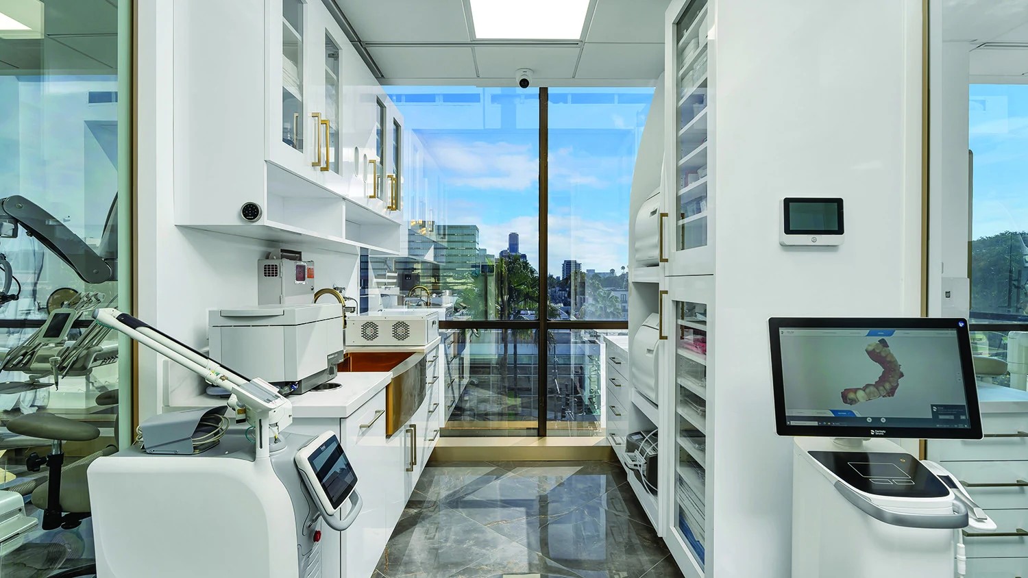
Microscope Dentistry
High-magnification dentistry using enhanced illumination and visualization to support precision in restorative care.
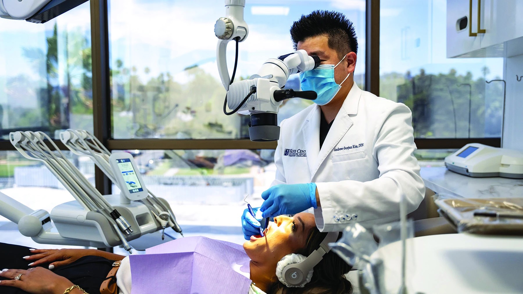
Magnification changes what the clinician can see.
A dental operating microscope provides magnification and focused illumination. In restorative dentistry, that can support detailed inspection of tooth structure, margins, cracks, existing restorations and operative fields.
- Enhanced illumination
- Magnified evaluation
- Restorative margin visualization
- Documentation and communication
- Precision-focused workflow
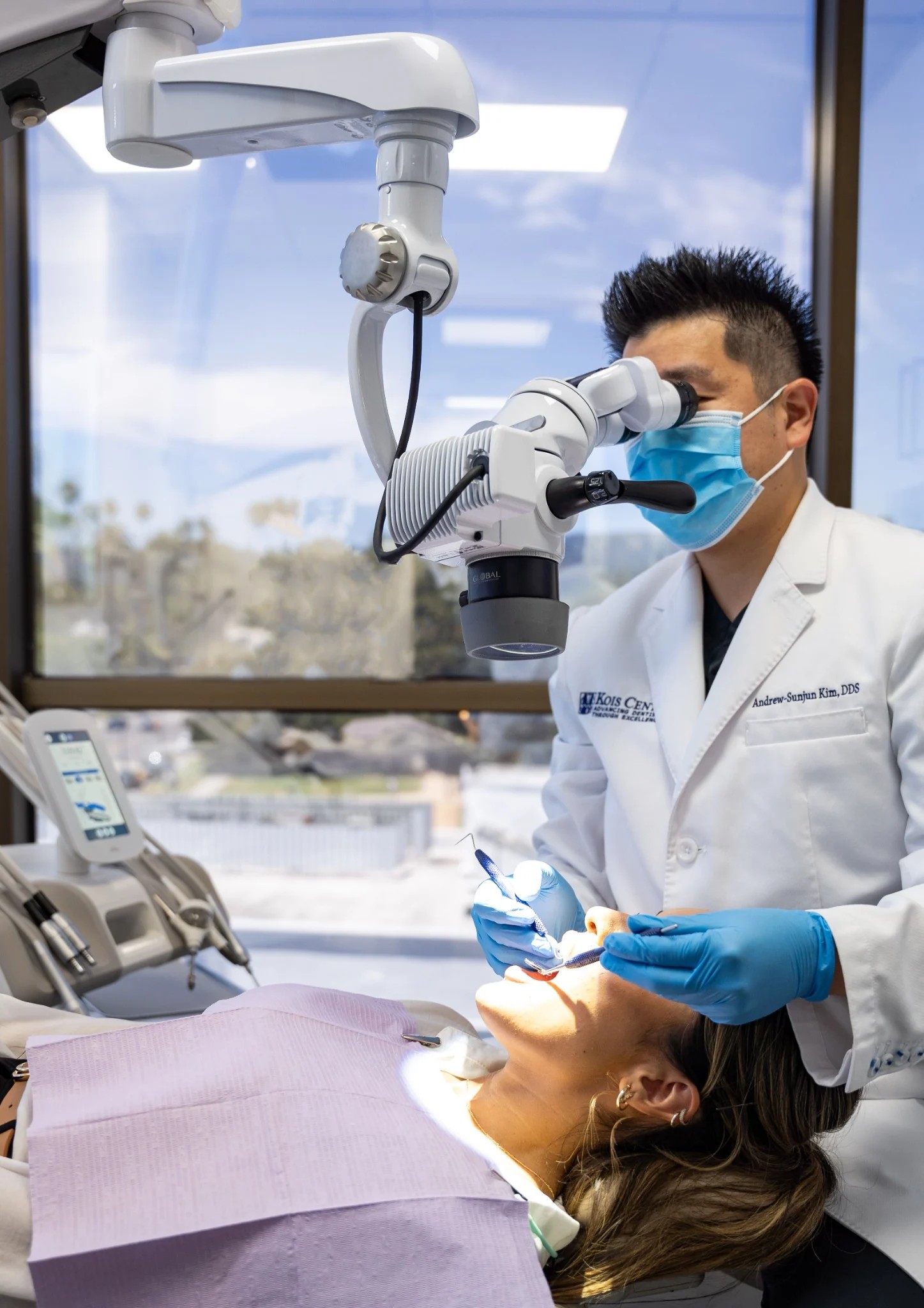
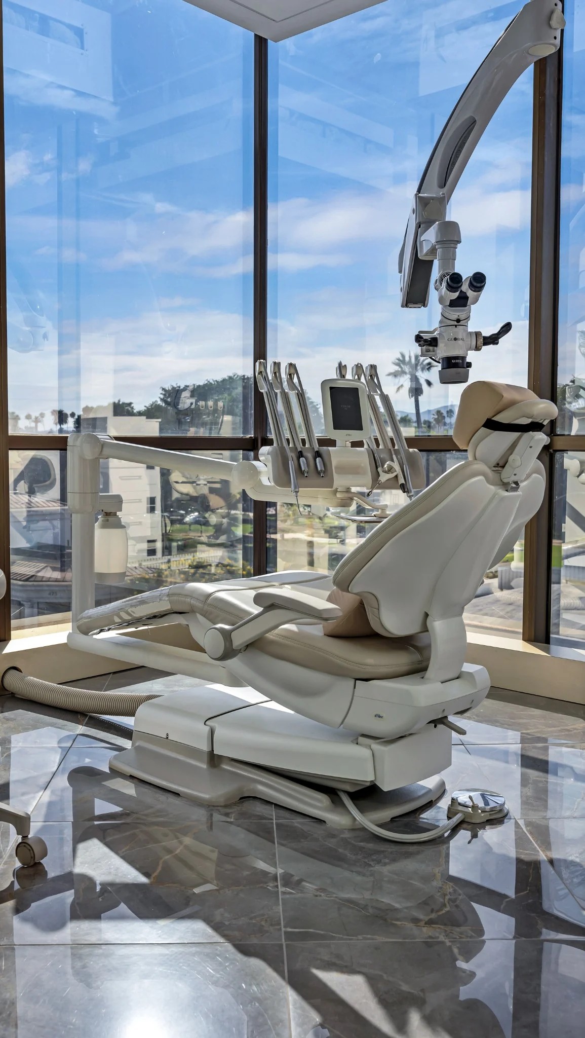
Seeing more does not automatically mean treating more.
Magnification is a diagnostic and execution aid. Clinical judgment remains essential to decide what findings are meaningful and what treatment, if any, is appropriate.
Microscope dentistry complements digital dentistry.
Microscopy, photography, scanning and 3D imaging answer different questions. Used together appropriately, they can improve visualization and communication.
What patients ask about microscope dentistry
Why use a dental microscope?
It provides magnification and illumination that can help the dentist inspect and work on small structures with greater visual detail.
Does microscope dentistry mean a procedure will be painless?
No. Magnification improves visualization; comfort is managed separately with anesthesia, technique and, when appropriate, sedation.
Is microscope dentistry used for all procedures?
It may be used selectively when enhanced visualization is useful.
A thoughtful plan starts with a thorough diagnosis.
See more. Work with finer detail.
The magazine emphasizes dental microscopy as part of Enzo Dentistry’s restorative and surgical workflows. Magnification and illumination can improve visualization of tooth structure, margins and fine clinical detail.